STONE project


The STONE project (Studies on T-ONE colorectal cancer) aims to identify biomarkers for lymph node metastasis in T1 CRCs in order to improve selection of patients for adjuvant surgery.


Please find background information about this project below. 

If you want to include a patient in this trial, click here:


  • Text Hover

Team and contact information


 Team

L.M.G. Moons, principal investigator

M.M. Lacle, principal investigator

Y. Backes, project leader


Phone Number

+31 6 1459 9001

+31 6 1887 3897

+31 6 1223 4744


E-mail

L.M.G.Moons@umcutrecht.nl

M.M.Lacle@umcutrecht.nl

Y.Backes@umcutrecht.nl

Background

The vast majority of patients with a T1 CRC do not have lymph node metastasis (LNM). T1 CRCs in the colon can potentially be removed with minimal invasive techniques such as endoscopic polypectomy or trans anal microsurgery (TEM). The benefits of treating patients with these minimal invasive techniques as compared to major surgery, are the low mortality rates (< 0.5% vs. 3%), minor complication rate (3% vs. 15%), and the less economic burden on national healthcare costs as the costs of a polypectomy are only 10% of the costs of major surgery.


The Problem

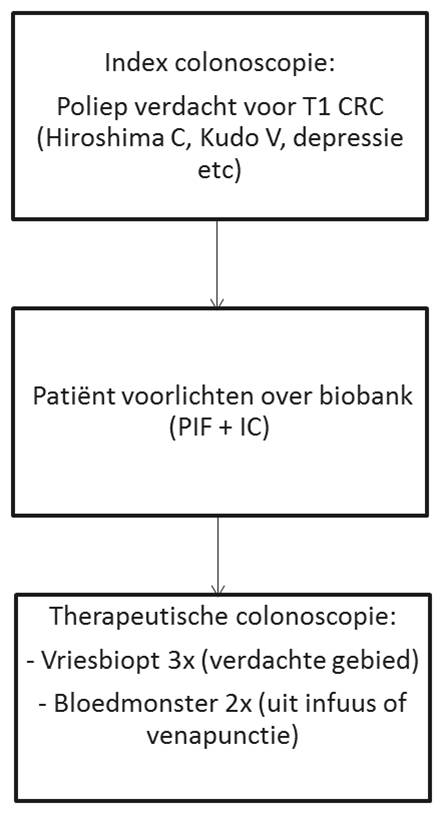
The difficulty is however to predict the presence of LNM prior to major surgery. Currently, only histologic markers are recognized to predict LNM. In the recently published Dutch guideline on treatment of CRC, the histological criteria grade of differentiation and the presence of lymph-angioinvasion have been stated as risk factors for LNM. In the absence of these criteria, the patient has an extremely low risk of LNM or recurrence of the CRC within the following 5 years. Only 25% percent of the T1 carcinomas can be recognized as low risk, and this group is advised to be treated with minimally invasive techniques. The remaining 75% of “high risk patients” are referred for major surgery. Of these, only 15% will be found to have LNM after surgery. The positive predictive value (PPV) of this approach is only 15% with a NPV of 100%. Therefore, a significant group of patients still undergoes major surgery without any benefit.


Solution

During the last decades, the insights in the mechanisms involved in the development of LNM have increased. This knowledge may be used to identify molecular markers indicative of the pathways involved in LNM. These markers may be used to predict the presence of LNM after minimal invasive resection of T1 CRC.

Goal / hypothesis

To obtain a panel of molecular predictors to predict the presence of LNM in T1 CRCs after resection with minimally invasive techniques. This set of biomarkers can then be used in daily practice to guide the decision which patients should be referred for adjuvant surgery. Based on the results in the past, we expect that our model will have an accuracy of 85%, and that at least 65% of T1 CRC patients can reliably be identified as low risk. This will decrease the number of unnecessary surgical procedures with 47%.

Design

The study consists of two separate parts which converge at the end of the study period.

  1. The first part concerns a biased approach based on all the markers detected in the review of the literature. A case control study on archival FFPE material will be performed. Cases and controls will be selected from the T1 CRCs working group database. All slides will be reviewed by expert pathologists. We will prioritize the candidate immunohistochemical markers based on the strength of each biomarker for predicting adverse patient outcome, and the variation in human marker expression (markers with very low variation have less chance of clinical impact). The most promising candidate markers will be tested by IHC on our T1 CRC patient set.
  2. The second part of the study concerns an unbiased approach with RNA sequencing of total and small RNA (miRNA, tRNA) to obtain information on the different pathways involved in LNM in T1 CRCs. Currently most data is obtained in stage II and III CRCs which often concerns TNM-stage T2-T3 tumors. It is not certain whether the results from gene profiling can be applied to T1 CRCs. Moreover, miRNA analysis has not been performed to evaluate signaling pathways for LNM in CRC. Snap frozen material will be prospectively collected from which RNA is isolated, from which different signatures will be analyzed.  

Inclusion and exclusion criteria

Inclusion:

  • 18 year or older
  • Colorectal polyp suspected for T1 CRC (Hirshima C, Kudo V, depressive area etc), as detected during screening, surveillance or diagnostic colonoscopy.


Exclusion:

  • No informed consent

Primary endpoint

Lymph node metastasis, as assessed by the pathologist

Study documents

Participating hospitals

We use cookies to ensure that we give you the best experience on our website.